Intravital Imaging
Headed by: Dr Max Nobis
e-mail: max.nobis@kuleuven.be
telephone: +32 16 713684
location: 08.159, O&N4, Campus Gasthuisberg
twitter: @_Intravital
ORCID: 0000-0002-1861-1390
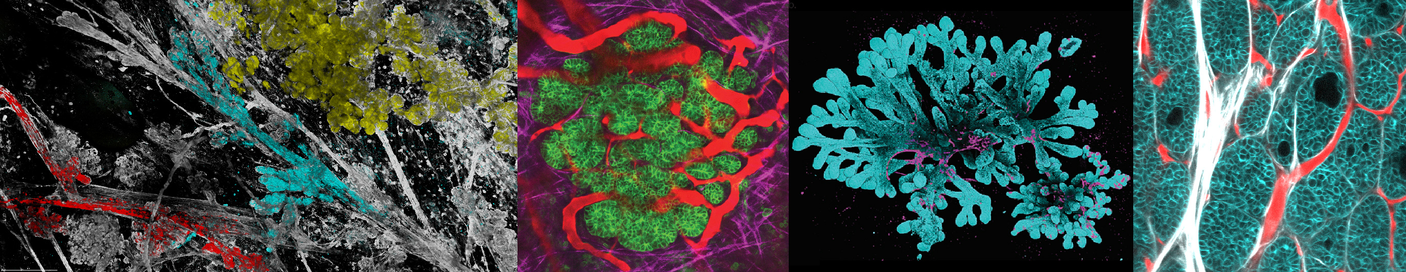
Tumors are highly dynamic ecosystems, in which the tumor cells and their micro-environment consisting of many different cell types, continuously evolve over time. To understand this intricate and complex interplay, it is crucial to study tumor evolution in space and over time, i.e. in 4 dimensions. With intravital microscopy single cells can be followed over multiple days to weeks in unperturbed in vivo tissues which makes intravital imaging the only method to study the phenotypic behavior and plasticity of individual tumor cells and wild-type cells in their natural niches. This technology has further been applied to study metabolic, neurologic, immunological interactions and infectious diseases.
The CCB Intravital Imaging Expertise Center harbors two high-end multiphoton microscopy systems dedicated to the in vivo imaging of cells and their native micro-environment. To this end a wide array of fluorophores, biosensors and in vivo imaging dyes can be supplied and recommended by the center. The setup of the imaging systems is specifically designed to allow deep in vivo tissue imaging in zebrafish and of both ventrally and dorsally localized tissues of interest in living mice. Both microscopes are specifically equipped with several accessories for long-term, multiday imaging sessions to follow tumor cell behavior over longer time periods. To gain optical access to the tissues of interest, small imaging windows have been developed and are utilized to visualize individual tumor cells in their native environment.
Services
- Design, planning and execution of intravital experiments
- Choosing and optimizing biosensor and/or dyes and stains from our collection
- Aiding in writing and review ECD applications
- Training on 2-photon microscope use
- Performing and training of surgical techniques (e.g. optical windows for various target organs, including but not limited to subcutaneous tumours, mammary gland/tumours, pancreas, liver, spleen, lung and brain)
- Tissue imaging ex vivo
- Customized tissue clearing and staining of intact tissue
- Single photon and 2-photon imaging of stains, tissue autofluorescence (e.g. SHG, collagen I)
- Tissue size can be in the cm range
- Post imaging processing
- 3D rendering of cleared tissues (e.g. tumours, whole organs)
- Quantification e.g. blood flow, extracellular matrix (ECM) content and organization
- Motion correction of intravital imaging
In house systems
Leica SP8 Location: O&N4 08.149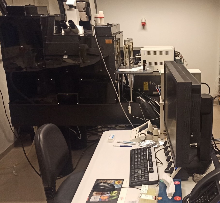
- upright system with heated chamber
- Single photon laser lines: 405, 458, 476, 488, 496, 514, 561, 633
- 4 internal HyD and 1 PMT detectors
- 2-photon lasers:
- 680-1060 tunable wavelength for 2P
- tunable OPO 1080-1300
- 4 external HyD detectors
- available filter cubes:
1. 460/50, 525/50, dichroic 495
2. 610/25, 736/128, dichroic 790
3. 525/50, 585/40, dichroic 560
4. 624/40, 685/40, dichroic 650
Leica SP8 DIVE Location: O&N4 09.339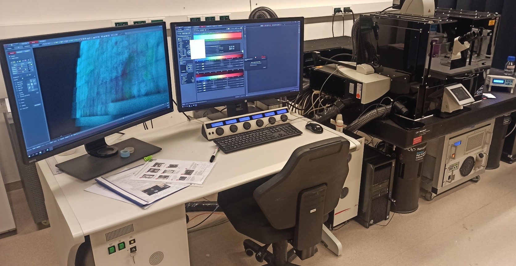
- Inverted system with heated chamber
- connected anesthesia unit
- Single photon laser lines: 405, 488, 552, 638
- 2 internal HyD and 1 PMT detectors
- 2-photon lasers:
- 1040 (fixed wavelength 2P)
- 680-1060 tunable wavelength for 2P
- 4 tunable external detectors (3 HyD, 1 PMT)
The CCB Intravital Microscopy Expertise Center strives to stay at the forefront of technology development. We continuously develop and invest in improved microscopy technologies, new imaging tools and refined surgeries for 3D and 4D in vivo imaging.
Current technologies trialed at the Center include:
Kratoscope (Kaer Labs) Period: 13th October 2022 -12th October 2023
Developed by Kaer Labs working in close collaboration with histology platform in Nantes University (France), the Kratoscope is an imaging device that can be 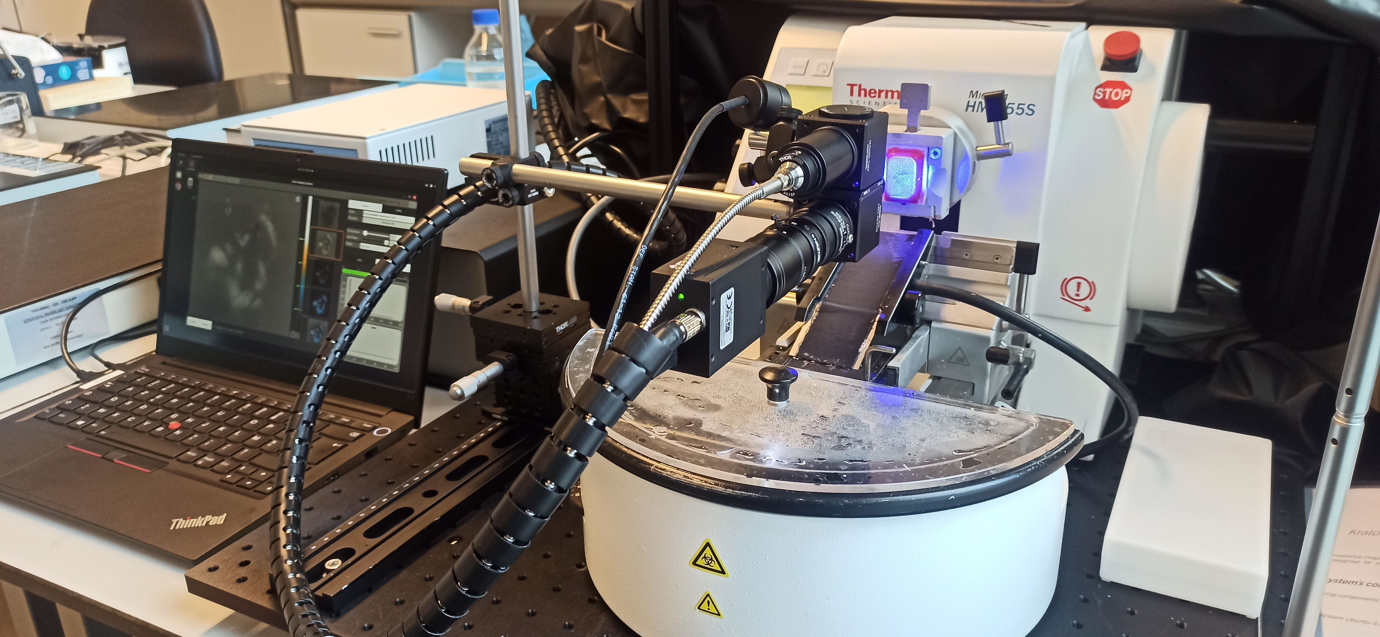 coupled to a conventional microtome to generate fast 3D reconstructions of any tissue in a semi- or even fully automated, and label-free way. The Kratoscope uses serial block face imaging, taking an image of the tissue block during the sectioning based on the autofluorescent properties of the tissue and collecting the signals at the range >500 mn. The resulting images can be rendered in a 3D histological reconstruction of the tissue (resolution up to ~3 µm). The tissue sections can be collected at the same time for further downstream processing, and this information can than later be integrated into the 3D tissue reconstruction.
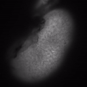
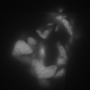
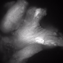
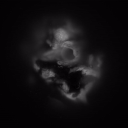
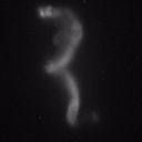
Example images of a kidney, pancreas, muscle tissue, thymus and fallopian tubes + ovaries:
More information can be found here: https://www.kaerlabs.com/3d-histology-kratoscope
Blaze Light Sheet Microscope (Miltenyi) Period: 28th November 2022 -12th December 2022
The UltraMicroscope Blaze is the only fully automated light sheet microscope for imaging large or multiple cleared samples at subcellular resolution. The system is equipped with a white light laser and the following filter options:
Excitation Filters Emission Filters
405/30 460/40
470/30 525/40
520/40 585/40
595/25 640/50
630/30 680/30
680/40 720/24
710/75 810/90
785/25
A standard sample chamber allows convenient imaging of multiple rodent organs or organoids. Additionally, the new XXL chamber is specially designed to provide the largest possible sample space for the UltraMicroscope Blaze. It enables fitting of samples as big as a human kidney or whole adult mouse models.
More information can be found here: https://www.miltenyibiotec.com/BE-en/products/ultramicroscope-blaze.html
Click below to go to the roadmap for the IVI Expertise Center.